-
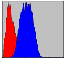
Figure 2: Flow cytometric analysis of Hela cells using CD133 mouse mAb (blue) and negative control (red).

Figure 1: Immunohistochemical analysis of paraffin-embedded human breast cancer tissues (left) and human esophageal cancer tissues (right) using CD133 mouse mAb with DAB staining.